Tooth-Saving Treatment Based on the Condition of Your Tooth
Not every painful or damaged tooth needs the same treatment. Your dentist will assess the pulp, roots, surrounding bone, existing restorations and overall condition of the tooth before recommending the most appropriate option.
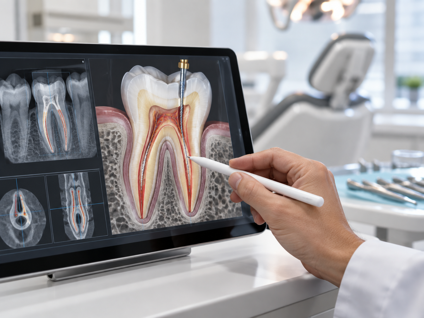
Remove Infection and Preserve a Restorable Tooth
Root canal treatment is used when the pulp inside a tooth becomes irreversibly inflamed, infected or damaged. The affected tissue is removed, the root canals are carefully cleaned and disinfected, and the internal space is sealed to help retain the natural tooth. Before treatment, we assess whether the tooth has enough healthy structure and support to be successfully restored afterwards.
- Treats infection or inflammation within the tooth
- Helps preserve a natural tooth when it remains restorable
- Addresses a common underlying cause of toothache and biting pain
- Avoids extraction when tooth preservation is clinically appropriate
- Coordinates root canal treatment with the final restoration
- Allows continued monitoring of the tooth and surrounding bone
Before & After
Illustrative only. Individual results may vary.
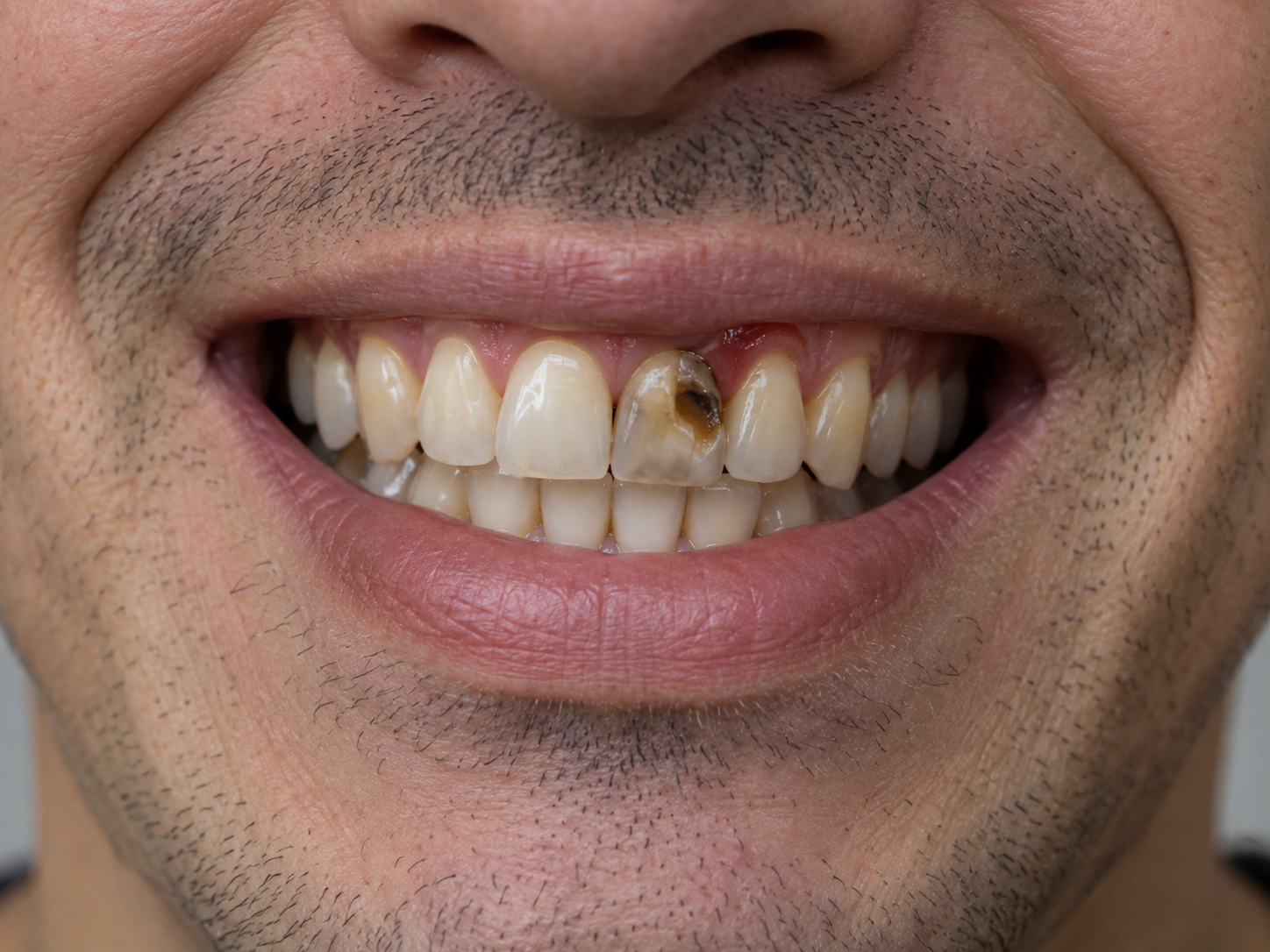
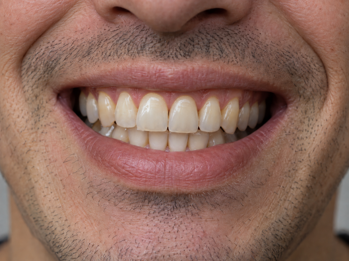
Illustrative image only. Root canal treatment outcomes vary between individuals and depend on factors including the extent of infection, tooth structure, root anatomy, cracks, periodontal support, final restoration and healing. Results cannot be guaranteed. A clinical examination and appropriate dental imaging are required to determine suitability and expected outcomes.
We examine the tooth, assess symptoms, perform pulp and bite tests and take appropriate dental X-rays. Additional imaging may be recommended when anatomy, previous treatment or surrounding structures require closer assessment.
Local anaesthesia is used for comfort. A dental dam is placed when appropriate to isolate the tooth from saliva and help maintain a clean treatment environment.
Inflamed or infected tissue is removed. The canals are measured, shaped and disinfected using appropriate endodontic instruments and solutions.
After cleaning, the prepared canal spaces are filled and sealed with endodontic materials designed for use inside the root canal system.
The tooth is restored with an appropriate filling, onlay or crown depending on how much healthy tooth structure remains. Follow-up evaluates symptoms, bite, restoration integrity and healing.
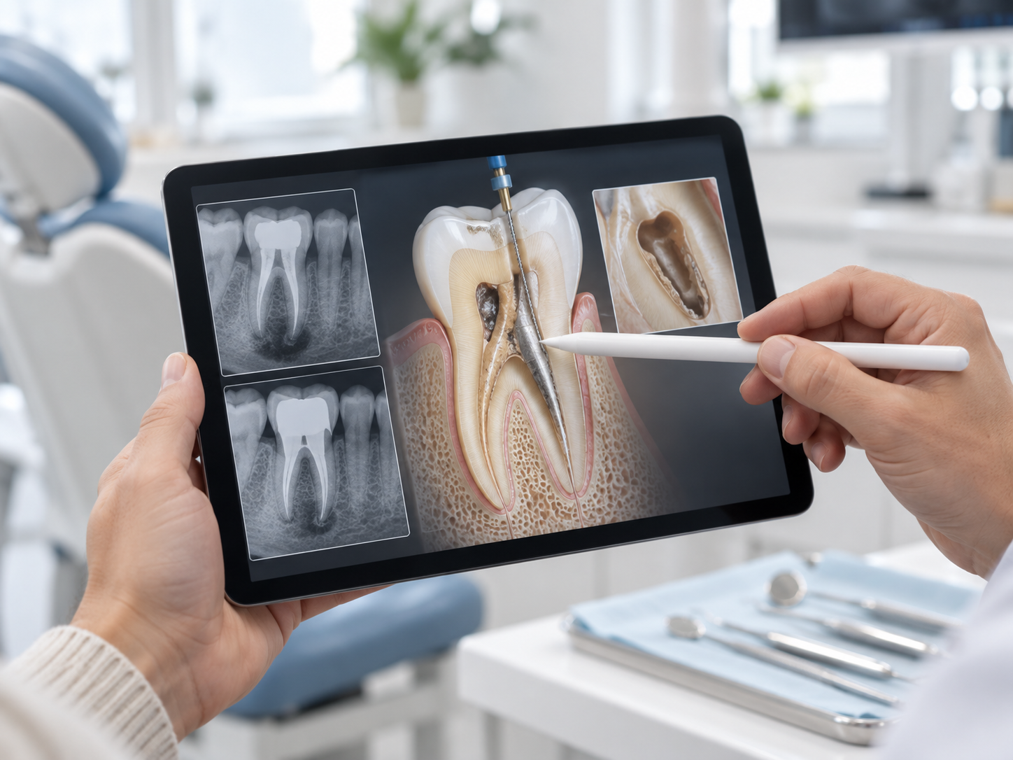
Give a Previously Treated Tooth Another Opportunity
A tooth that has already received root canal treatment can sometimes develop new symptoms or persistent disease. This may occur because of missed anatomy, leakage around a restoration, new decay, difficult canal anatomy or another problem affecting the tooth. Root canal retreatment involves reopening the tooth, removing previous root-filling materials and disinfecting the canal system again. Before retreatment, we assess whether the tooth is restorable and whether retreatment, apicoectomy or extraction is the most appropriate approach.
- Offers another opportunity to preserve a restorable tooth
- Allows previously treated canals to be reassessed
- May address missed canals or renewed bacterial contamination
- Can be considered before root-end surgery in appropriate cases
- Coordinates treatment with the condition of the existing crown or restoration
- Allows prognosis and alternative options to be discussed before treatment
Before & After
Illustrative only. Individual results may vary.
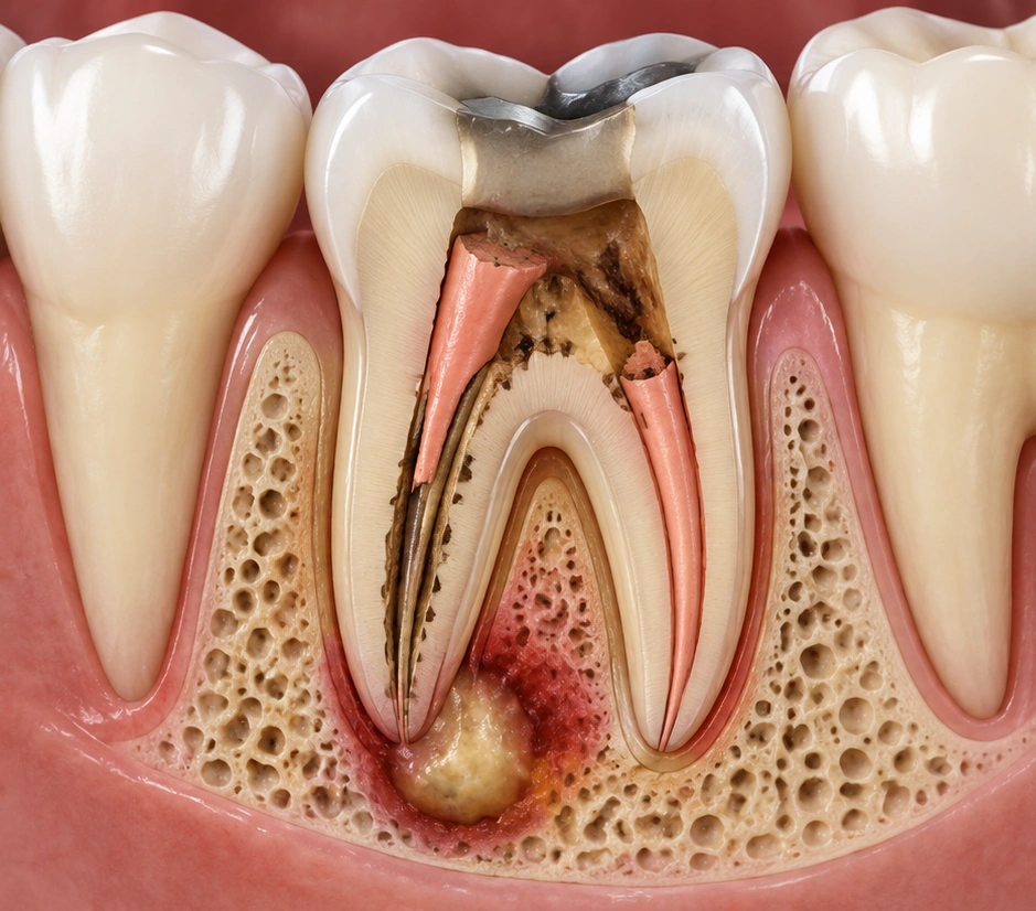
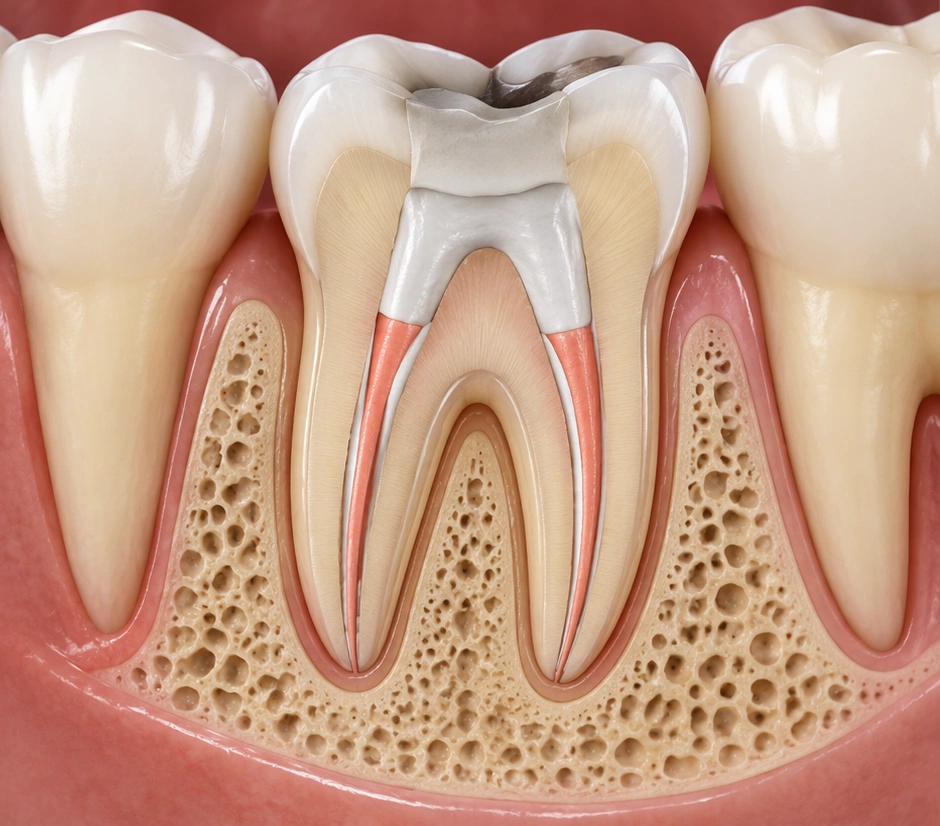
Illustrative image only. Root canal retreatment outcomes vary between individuals and depend on factors including the condition of the previously treated tooth, root canal anatomy, infection, cracks, restoration quality, periodontal support and healing response. Results cannot be guaranteed. A clinical examination and appropriate dental imaging are required to determine suitability and expected outcomes.
We review your symptoms, previous treatment, existing restoration, gum and bone support, dental imaging and the risk of cracks or fracture.
After local anaesthesia and isolation, access is created through the existing restoration. A crown, post or core is altered or removed only when clinically necessary.
Existing canal materials are removed so that the canal system can be inspected and reached again.
Accessible canals are cleaned and disinfected before being filled again. If part of the disease cannot be managed through the canal system, root-end surgery may still be considered.
A secure final restoration is arranged and follow-up reviews symptoms, function and changes around the root.
Treat Persistent Infection Around the Root Tip
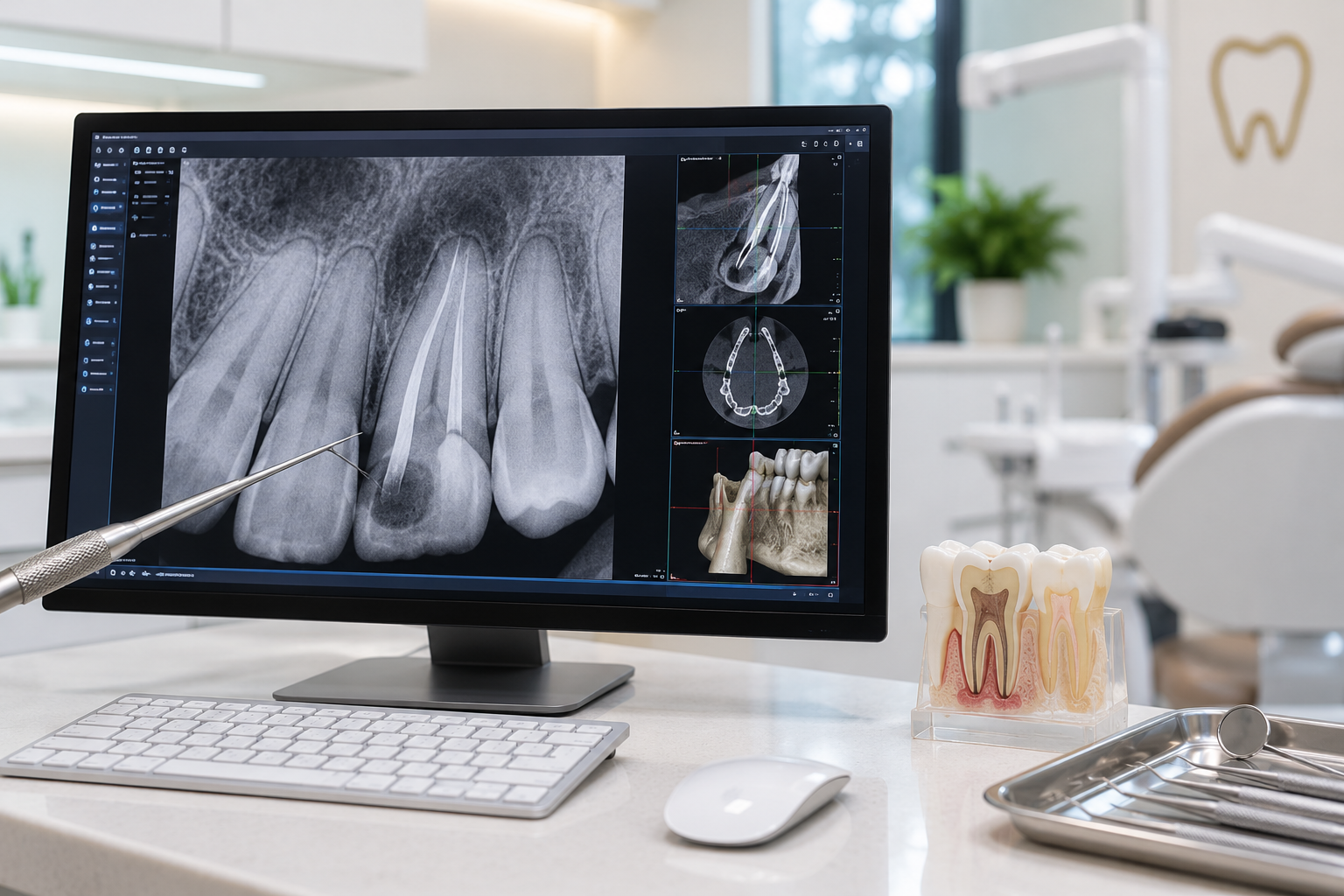
An apicoectomy is a minor root-end surgical procedure that may be considered when infection or inflammation persists around the tip of a previously root-treated tooth and conventional retreatment is not appropriate or has limited benefit. The affected area is reached through the gum, diseased tissue is removed and the end of the root is treated and sealed when indicated. Before recommending surgery, we compare root canal retreatment, apicoectomy and extraction based on the condition and long-term outlook of the tooth.
- Treats persistent disease located around the root tip
- May help retain a strong and restorable tooth
- Provides direct access to the affected root-end area
- Allows surrounding inflamed tissue to be removed
- Can be considered where conventional retreatment is impractical
- Follow-up imaging can be used to assess bone healing
Before & After
Illustrative only. Individual results may vary.
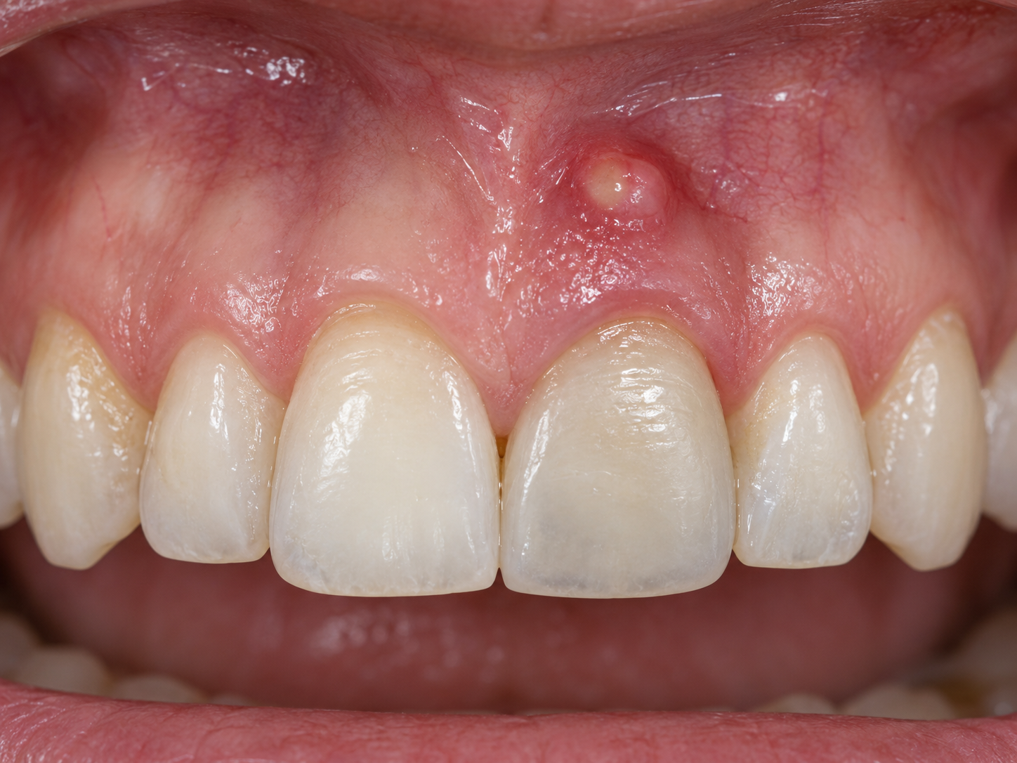
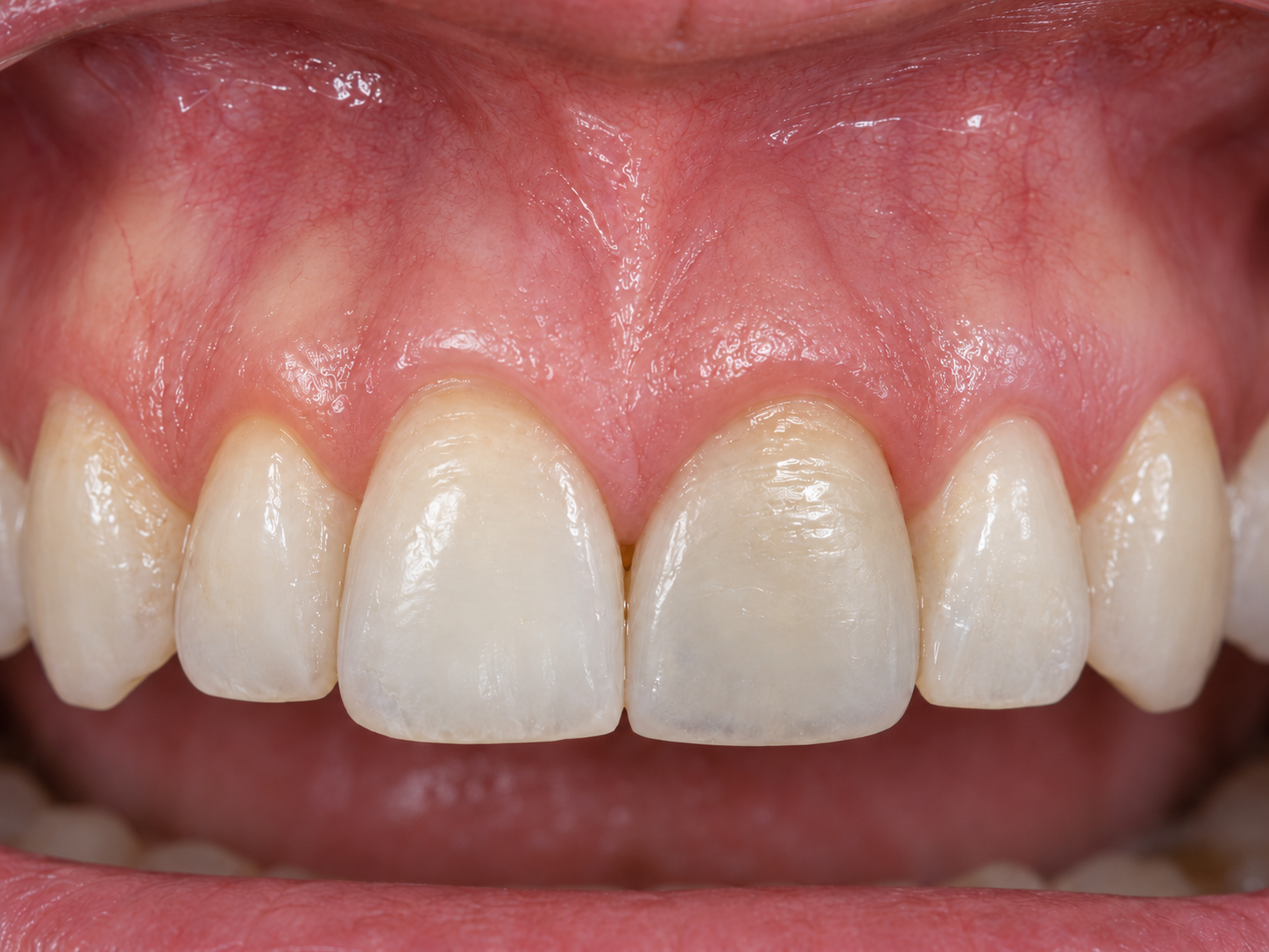
Illustrative image only. Apicoectomy outcomes vary between individuals and depend on factors including the condition of the root-treated tooth, extent of infection, root anatomy, bone and gum health, restoration quality and healing response. Results cannot be guaranteed. A clinical examination and appropriate dental imaging are required to determine suitability and expected outcomes.
We review the existing root filling, crown or restoration, root anatomy, cracks, periodontal support and imaging. Alternative treatment options are discussed before surgery is recommended.
Local anaesthesia is used. A small surgical opening through the gum and surrounding bone provides access to the affected root tip.
Inflamed or infected tissue around the root is removed and the root end is carefully inspected.
A small portion of the root tip may be removed and the root end is prepared and sealed with an appropriate biocompatible material when required.
The area is sutured and post-operative instructions are provided. Follow-up evaluates comfort, gum healing and later radiographic bone healing.
Preserve Healthy Living Pulp When the Tooth Allows
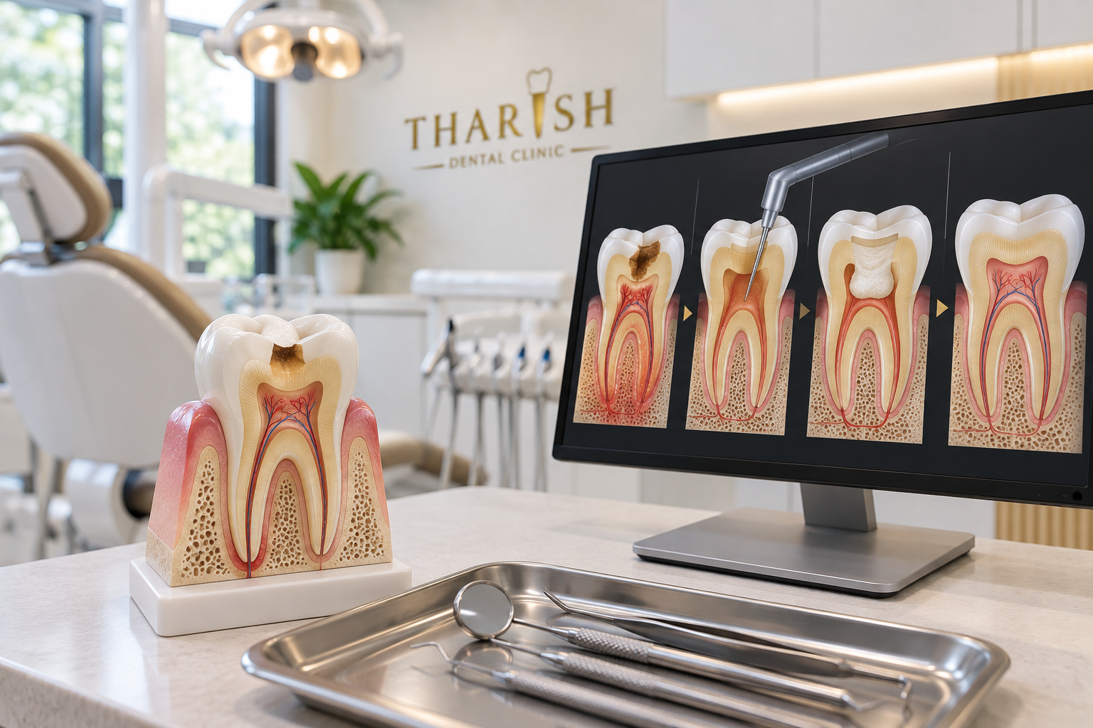
Deep decay or dental trauma does not always mean that all of the pulp inside a tooth must be removed. Vital pulp therapy aims to preserve healthy living pulp tissue when the clinical findings indicate that healing is possible. Depending on the condition of the tooth, treatment may involve protecting exposed pulp tissue or removing only the inflamed portion while preserving healthy tissue deeper inside the tooth. This approach can be especially valuable in young permanent teeth that are still developing.
- Preserves living pulp tissue when clinical conditions are favourable
- Can support continued root development in young permanent teeth
- May avoid full root canal treatment in selected cases
- Removes diseased tissue while retaining healthy pulp where possible
- Uses a well-sealed final restoration to protect the treated area
- Includes ongoing monitoring to detect changes early
Before & After
Illustrative only. Individual results may vary.
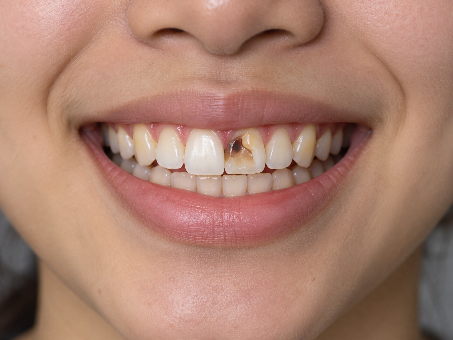
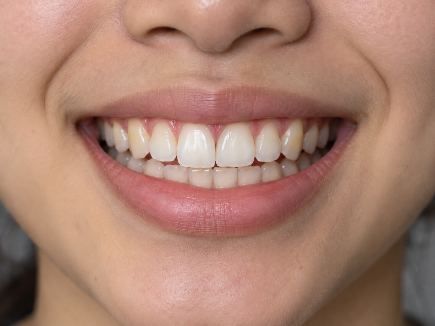
Illustrative image only. Vital pulp therapy outcomes vary between individuals and depend on factors including the extent of decay or trauma, pulp condition, tooth development, restoration quality, oral hygiene and healing response. Results cannot be guaranteed. A clinical examination, pulp testing and appropriate dental imaging are required to determine suitability and expected outcomes.
Your symptoms, tooth history, clinical examination, pulp tests and imaging help determine whether healthy pulp tissue may be preserved.
Local anaesthesia and dental isolation help provide comfort and reduce contamination during treatment.
Decay and unhealthy tissue are carefully removed while preserving recoverable pulp whenever clinical findings allow.
An appropriate biocompatible material is placed over the pulp tissue and the tooth is sealed with a durable restoration.
Follow-up appointments reassess symptoms, pulp response, restoration integrity and radiographic development.